There is nothing like seeing something brand new that has never been seen before down the microscope.
Here are some of my own microscopy images that shaped my research career and are pretty to look at.
A “POSH” PhD
My PhD in Cell Biology instilled in me the joy of seeing for myself the tiny world inside us.
left = POSH (Plenty of SH3s) protein the DNA for which was injected into the cell; right = red-labelled transferrin that the cells are trying to take-up (this is an iron-binding protein); white bar = 30 micro-metres (pretty small!).
Meet POSH, which stands for Plenty Of SH3s (shown on the left). This was the protein that took up 4 years of my life as the subject of my post-graduate PhD cancer research studies. I love this particular picture as it shows the strange shapes that POSH makes inside cells in a dish when you inject DNA to make lots of POSH, and tag it so that you can see it. We never did solve the mystery of why/how it does this.
Do you see how being full of POSH means that the cell cannot take up the protein called Transferrin seen on the right panel? Here the transferrin protein was linked to a fluorescent dye so I could add it to the cells after I injected them with the POSH DNA and see it under the microscope.
We found that POSH is able to stick to and block the function of a protein (dynamin) that’s a gate-keeper for what gets inside mammalian cells. We could never prove it does this when we don’t artificially put loads of POSH into a cell, so these pretty pictures remain published only in my PhD thesis.
Salmonella on or inside human cells
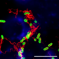
Salmonella Typhi bacteria on the outside of human cells in a dish.
The bacteria were prevented from invading the cells by a chemical (cytochalasin D) that inhibits changes in the actin cell skeleton (also termed the cytoskeleton) needed for bacteria to invade and get inside.
green = Salmonella Typhi (strain BRD948 Ty2 delta aroC aroD htrA – this is a safe vaccine candiate strain) surface sugars (Vi capsule); red = Salmonella Typhi tails (flagella Hd antigen); blue = human actin cytoskeleton; white bar = 10 micro-metres (so you can see the bacteria are very small, hence calling them microbes!).
This is Salmonella enteric subspecies I serovar Typhi, or S. Typhi as we often call it. This bacterium causes typhoid fever. Here the sugars on its surface are shown in green and its protein tails (flagella) are in red and it’s in the process of invading some human cancer cells in a dish (here blue is the human cell protein actin).
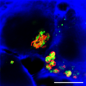
Salmonella Typhi bacteria inside cells. The ones still on the outside were killed with the antibiotic gentamicin and washed away, so only the inside ones are visible here. Staining details as above.
After the Salmonella Typhi bacteria have gone inside they have their flagella wrapped around them, like they are all squashed up, which indeed they are, as they are inside intracellular vesicles (like bags surrounded by a fatty layer) within the cells.
These two pictures were taken with a confocal microscope that uses lasers to make a precise plane through the sample visible, so you get this kind of cross-section. These images were never published, but were a clinching factor during my interview for my next job with Andrew Camilli’s team in Boston, MA, USA, as he thought they were impressive. I think they’re fascinating and beautiful.
The cells used here are INT 407 (HeLa derivatives), derived from a human cervial cancer (cancer cells are easy to keep growing in dishes, unlike most normal human cells). To learn more about how HeLa cells were derived from a cancer without the patient’s consent when she died from cancer in 1951, which is not allowed today, but have facilitated a lot of science done since, you can read “The Immortal Life of Henrietta Lacks”, reviewed here.
